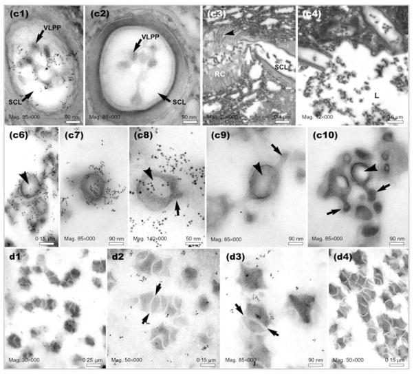
Fig. 5.
Electron micrographs of sections prepared from positions C and D of the L. heterotoma long gland and reservoir, respectively, as indicated in Fig. 1(d). Panels c1 and c2 show precursors of VLPs within the smooth canals (transverse sections). Smooth canals (SC) appear to be connected directly to rough canals (RC) (see white arrow in panel c3). The smooth canal empties into the long gland lumen (L) (panel c4). Panels c6–c10 show VLPs in the long gland lumen at position C. Some VLPs are spiked (arrows) and have acentric electron-light regions inside the VLP body (arrowheads). High levels of p40 are associated with developing VLPs (panels c6–c8). Panels d1–d4 show VLPs in the reservoir at position D (see Fig. 1d). Arrows show the presence of ‘tracks’ separating maturing VLPs. Panels c2, c9 and d4 are negative controls (primary antibody omitted), whereas samples in panels c3, c4 and c10 were not immuno-stained.

