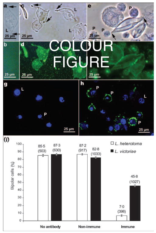
Fig. 7.
(a–h) Detection of p40 and p47.5 proteins by indirect immunofluorescence using anti-p40 antibodies. All haemocytes used were from hopTum-l/Y larvae and were treated with either L. heterotoma (a–d, g–h), or L. victoriae (e, f) fluid. For the negative controls (a, b), primary antibody was omitted. (a, c, e) Phase view of cells in (b), (d) and (f), where anti-p40 antibodies were visualized by FITC-linked secondary antibody. Arrows point to the tapering ends of the lamellocytes as they assume bipolar morphology prior to lysis. Untreated lamellocytes do not become bipolar (not shown). (g, h) Confocal images showing the association of hopTum-l haemocytes (counterstained with the nuclear dye TOTO-3) with anti-p40 antibody within plasmatocytes treated with L. heterotoma fluid (h) and the negative control (g). P, Plasmatocyte; L, lamellocyte. (i) Inhibition of formation of bipolar cells by anti-p40 antibodies. The percentage of lamellocytes that became bipolar cells is noted on top of each bar. The total number of lamellocytes examined is indicated in parentheses. Non-immune: mouse serum was used as a negative control; immune: anti-p40 antibody-treated fluid from L. heterotoma or L. victoriae. Suppression of bipolar cell formation by the anti-p40 antibody was highly significant (z>16, P<0·003) in pairwise z tests for independent proportions between the no antibody/non-immune (negative controls) and the immune (experimental) in vitro assays with fluid from either wasp.

