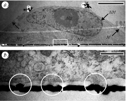
Figure 5.
Typical TEM images of HEK cells grown on smooth substrates. (a) This image shows a HEK cell grown for 4 days on a PLL-coated substrate and (b) in higher magnification (white rectangle in (a)). The cell and prominent cellular structures were well preserved: membrane (M); nucleus (N) with nucleolus (Nu); nuclear membrane (NM); and mitochondria (Mi). The cell attached tightly to the substrate surface, which is marked by the gold-sputtered layer (black line). Differences were found in the membrane attachment: areas of close adhesion (white circles) and areas with enhanced distance. Scale bars, (a) 5 μm, (b) 1 μm.

