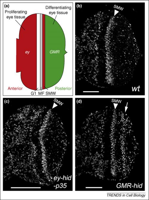
Figure 3.
Two modes of apoptosis-induced compensatory proliferation in proliferating and differentiating eye tissues in Drosophila. (a) Schematic outline of the late-third instar eye imaginal disc. Anterior is to the left. The developing larval eye disc is composed of the anterior proliferating tissue (red) and the posterior differentiating tissue (green), which are separated by G1-arrested cells, the morphogenetic furrow (MF) and the second mitotic wave (SMW). The eyeless (ey) promoter is expressed in the anterior part (red); the GMR promoter is expressed in the posterior part (green) and in the SMW. (b) An eye imaginal disc from a 3rd instar wild-type larva labeled with BrdU as the proliferation marker. The anterior half of the disc [red in (a)] is heavily proliferating. Posterior to the SMW [green in (a)], proliferation has stopped and cells are differentiating into photoreceptor neurons and accessory cell types. The SMW is marked by a white arrowhead. (c) Co-expression of hid and P35 under ey control (ey-Gal4, UAS-hid, UAS-P35) triggers overgrowth of the anterior compartment [red in (a); compare with (b)]. The white bar indicates the extent of overgrowth [compare to (b)]. (d) Expression of hid under GMR control (GMR-hid) triggers compensatory proliferation (white arrow) posterior to the SMW [green in (a); compare with (b)]. Part (a) adapted, with permission, from Ref. [13].

