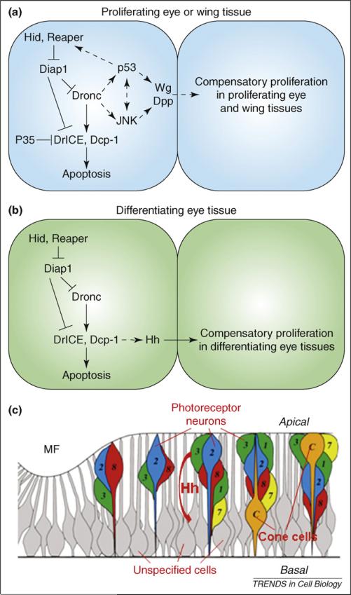
Figure 4.
Models of distinct mechanisms of compensatory proliferation in proliferating versus differentiating tissues in Drosophila. (a,b) Molecular mechanisms of apoptosis-induced compensatory proliferation in proliferating tissues (a) and differentiating tissues (b). An apoptotic cell (left) and a cell that is induced to undergo compensatory proliferation (right) are shown. Dashed arrows indicate unknown interactions. The scaffolding protein Ark is omitted for clarity (Figure 2). (c) Schematic outline of various cell types in the differentiating eye tissue. Hh produced in apical dying photoreceptor neurons (numbered colored cells) non-autonomously induces cell-cycle re-entry of basally-located unspecified cells (gray cells). ‘Apical’ and ‘basal’ refer to the apical and basal sides of the disc. The SMW is omitted for clarity. Part (c) adapted, with permission, from Ref. [13].

