
Figure 1. Intranasal application of VSV induces a vigorous mixed cellular infiltrate in the brain.
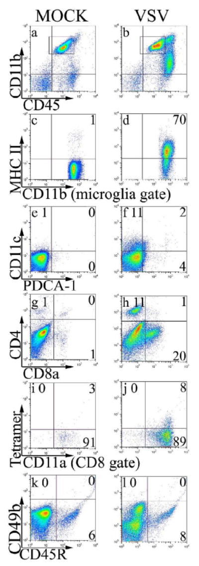
Mice were given either PBS (Mock) or intranasal VSV at 2×10⁵ PFU (VSV). Eight days post-infection, leukocytes were isolated from the brain and the infiltrate characterized by flow cytometry. Microglia and infiltrating leukocytes were first identified by forward and side scatter profiles. Within this gate microglia were defined as CD11b+ and CD45low/int (box in panels a-b) and expression of MHC class II was evaluated on microglia-gated cells (panels c-d). To characterize other infiltrating cell types, we assessed gated leukocytes (forward and side scatter gate) for DCs (panels e-f) and T cell subsets (panels g-h). To identify VSV-N T cells, co-expression of CD11a and tetramers were assessed on gated CD8+ cells (panels i-j). NK cells and B cells were identified in the leukocyte gate as CD45highCD49b+ and CD45high CD45R+, respectively (panels k-l). This data is derived from the pooled brains of 4 mice per group.