
Figure 6. Systemic ablation of peripheral dendritic cells does not suppress the virus-induced IFN-γ response in the CNS.
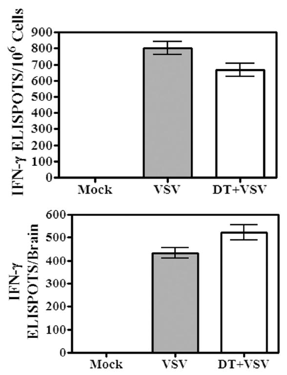
Mice were given a single intranasal instillation of VSV after being treated with either PBS (VSV) or DT (DT + VSV). Control mice were not infected (mock). Six days post-infection, brains were removed, pooled and leukocytes isolated by Percoll gradient centrifugation. Cells were seeded into ELISPOT plates in triplicate at up to 2×10⁶ cells/well and incubated overnight. No exogenous virus or viral peptide was added to these cultures. The following day plates were developed and the number of ELISPOTs/input cell number determined under a dissecting microscope. The number of ELISPOTS/106 cells determined for each triplicate input cell number was averaged and expressed as the mean ± S.E.M. (top panel). The total number of IFN-γ-producing cells/brain was then calculated based on this value and cell recoveries (bottom panel) per organ. Brains from 4-5 mice were pooled within each group. This experiment is representative of two additional experiments.