
Fig. 1.
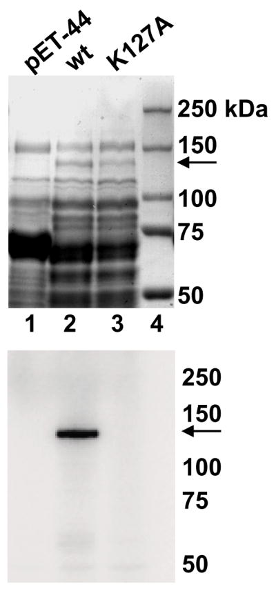
The guanylyltransferase domain of TbCgm1 binds GMP. Soluble protein lysates from bacteria expressing the NusA protein (pET-44, lane 1), amino acids 1 - 785 of TbCgm1 fused to the NusA protein (wt, lane 2), and a fusion protein with the indicated mutation (K127A, lane 3) were fractionated by SDS-PAGE and stained with Coomassie blue dye (upper panel) or tested for GMP binding (lower panel, an autoradiograph is shown). The sizes (in kilodaltons) of marker proteins are shown on the right and the position of the NusA-TbCgm1 fusion proteins is indicated by an arrow.