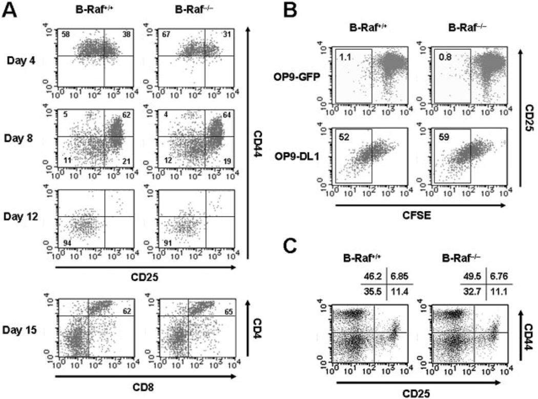
Figure 2. Pre-TCR-mediated β-selection is not affected by B-Raf deficiency.
(A) Phenotypic changes of DN cells differentiated from wild-type and B-Raf−/− FL-derived progenitors in co-culture with OP9-DL1 for various times. Developmental progression of the T cell lineage was assessed on days 4, 8 and 12 by CD25 and CD44 expression profiles on CD4−CD8− DN-gated cells. In the lowest panels, percentages of CD4+CD8+ DP cells co-cultured with OP9-DL1 for 15 days are indicated. Lymphoid cells were gated based on their SSC and FSC profiles. (B) DN T cell progenitors derived from B-Raf+/+ and B-Raf−/− FL cells co-cultured with OP9-DL1 for 8 days were loaded with CFSE and re-seeded on OP9 GFP (upper panels) or OP9 DL1 (lower panels). After 48 h, proliferation indicated by CFSE dilution and CD25 expression on DN cells during β-selection were determined. Dot plots show cells gated to eliminate CD4+CD8+ DP and OP9-DL1 cells. (C) Thymocytes from B-Raf+/+ and B-Raf−/− chimeric mice 7 wk after transfer of FL cells into RAG2−/− mice were isolated by positively sorting with anti-Thy-1 magnetic beads. The plots for CD44 and CD25 expression were gated on the CD4−CD8− DN population. The data are representative of three animals per group in two independent experiments with similar results, and the percentages of gated cells that fall into each quadrant are shown.

