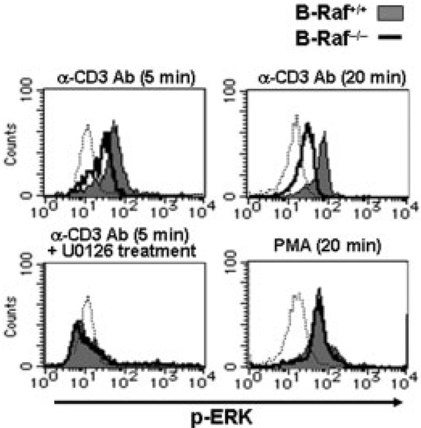
Figure 5.
Total thymocytes were stimulated by crosslinking of CD3 with 10 µg/mL anti-CD3 Ab and anti-hamster Ig Ab for 5 or 20 min (upper panels) or by incubation with 25 ng/mL PMA for 20 min (lower right panel). Cells were treated for 30 min with 10 µM U0126 prior to TCR stimulation (lower left panel). Histograms show profiles of phospho-ERK in B-Raf+/+ (filled gray) and B-Raf−/− (bold line) cells gated on CD4+CD8+ DP cells from DN cell-chimeric mice. Dotted lines indicate the status of phospho-ERK in unstimulated B-Raf+/+ DP cells. The data are from one representative of three independent experiments.

