Figure 2.
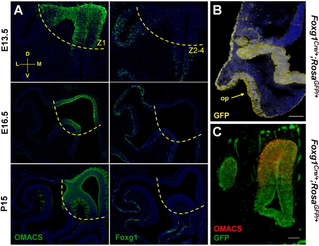
Foxg1 becomes restricted to ventrolateral regions of the olfactory epithelium during development. A, Time course of Foxg1 and OMACS immunohistochemistry. In coronal sections through the olfactory epithelium, there is complementary expression of the dorsomedial (Z1) marker OMACS and Foxg1 in the ventrolateral zones (Z2–4). B, Transverse section of an E10.5 Foxg1-Cre;Rosa-GFP mouse demonstrates that cells of the olfactory placode (arrow), telencephalon, nasal retina, lens, and surface ectoderm are derived from Foxg1-expressing cells. C, Using the same lineage tracing strategy coupled with an anti-OMACS antibody, it is clear that although Foxg1 becomes downregulated dorsally (A), the dorsal tissue did express Foxg1 at an earlier time in development. A coronal section at E13.5 with dorsal to the top and medial to the right is shown. Scale bars: B, 100 μm; C, 50 μm.

