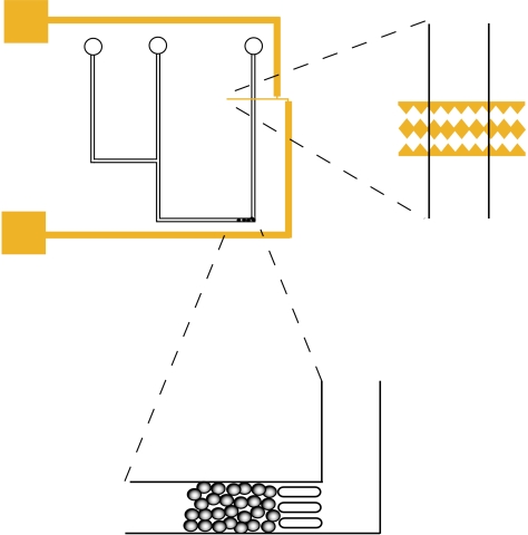
Figure 1.
Schematic showing the saw-tooth gold electrodes (on a glass slide) positioned within the PDMS microchannels, and the bead bed for protein capture created by trapping microspheres with three approximately 25 μm PDMS pillars. Simple microchannels of depth approximately 50 μm and width approximately 100 μm were used, with two inlets (one for the cell suspension and one for introducing the microspheres and/or a buffer for flushing through the system) and a single outlet.

