Figure 4.
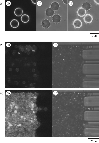
(a) Confocal images of the anti-β-actin modified beads, some of which have been incubated with cell lysate to demonstrate molecular capture of GFP–β-actin. (i)–(iii) The fluorescence image (excited at 488 nm and emission collected above 505 nm), transmitted light image and an overlay of the fluorescence and transmitted light images. The non-fluorescent beads have not been incubated with cell lysate, whereas the beads that were incubated with cell lysate show a clear fluorescence signal. (b) Anti-actin modified beads loaded into a microfluidic channel and trapped by rectangular approximately 25 μm PDMS pillars. (c) Fluorescence confocal image showing the capture of GFP–β-actin from a lysed cell population as it flowed over the bead bed.

