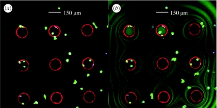
Figure 4.
(a) An LSM image of unsealed microwells showing the presence of the deposited Pt-porphyrin microspheres and healthy mouse macrophage stained with Calcein AM live stain in green. (b) An LSM image of sealed microwells showing the deposited sensors and the seeded macrophages. The symmetric wavy lines are indicative of an adequately sealed system.

