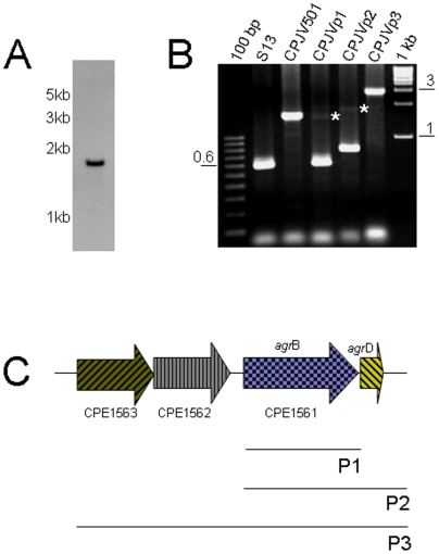
Figure 3. Generation of a C. perfringens agrB mutant and complementing strains.
A) Southern blot analyses, as described in Fig. 2, using EcoRI-digested DNA from CPJV501 and a Dig-labeled probe that detected a single copy of the erm gene. Size of DNA fragments, in kilobases (kb) is shown at left. B) PCR was performed with DNA extracted from the indicated strain and the following pair of primers, agrBFwd and agrBRev in reactions containing DNA from strain 13 (S13), CPJV501 and CPJVp1; agrBFwd and argDR for CPJVp2 and agrF1 and agrD100R for CPJVp3. DNA ladders (100 bp or 1 kb) were included in the first and last lane of the gel. Asterisks show the expected PCR product when the primers amplified the Tn5-disprupted agrB gene. C) Genes cloned in the E. coli-C. perfringens shuttle plasmid pJIR750 to complement the agrB transposon mutant. As shown, P1 encodes the agrB gene alone, P2 the agrB and agrD genes and P3 encodes two-genes (CPE1562 and CPE1563) upstream the agrB gene (CPE1561) and agrB and agrD.

