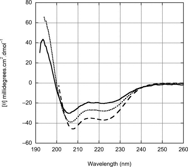
Figure 4.
Concentration dependent circular dichroism spectra of httNT. (A) httNT in aqueous buffer (see Methods) at 35 °C in concentrations of 3.8 μM (———),7.5 μM (······), and 18.9 μM (------). ContinLL 58 predicts significant secondary structure: 12% unordered, 4% β-strand, 20% turn, 8% polyproline type II helix, and 55% α-helix. (B) httNT in the presence of 10% trifluoroethanol (TFE) at 37 °C in concentrations of 7.5 μM (———), 18.9 μM (······), 94 μM (------). In the presence of this relatively low TFE concentration, the httNT adopts an α-helical structure, as evidenced by the negative bands at 208 and 222nm. The development of structure is protein concentration dependent, suggesting an oligomeric state under these conditions.


