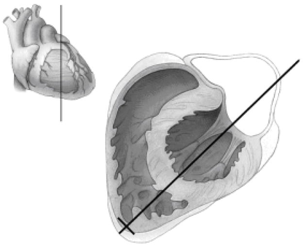
Fig. 4.

Illustration of Step 2. The long black line from the cardiac apex through the mitral valve defines the plane of a 4-chamber view. The volume sketch in the upper left-hand corner shows the plane of the main illustration in relation to the surface of the heart.
