
Figure 6.
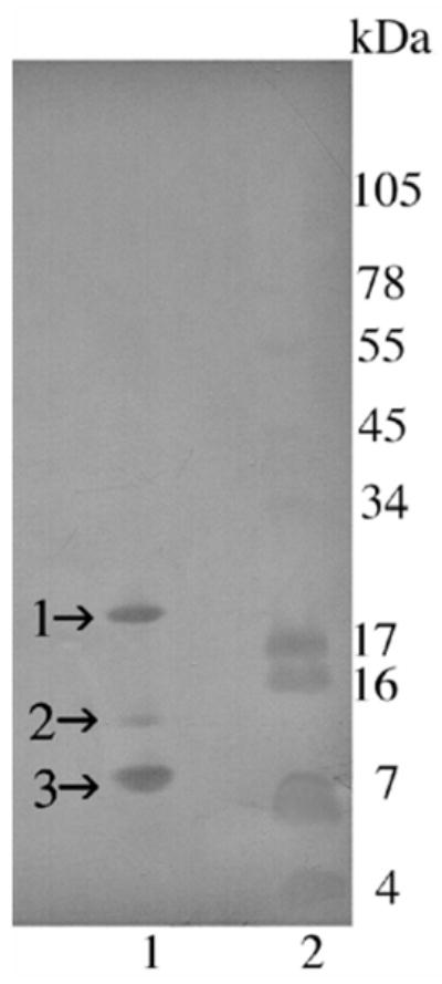
Transferblot of SDS-PAGE of C. o. helleri cationic exchange fraction V (hellerase) (Fig. 4B). A total of 5 μg of venom fractions were run on a 10–20% Tricine gel. The gel was over laid on an Immobilon-p transfer membrane and proteins were transferred using a Bio-Rad Trans-Blot Semi-Dry transfer cell at 100 mA for 1 h. Lane 1: Fraction FV collected without EDTA and run under non-reducing conditions; Lane 2: SeeBlue Plus2 Markers (Invitrogen™). The membrane was stained with 0.02% Coomassie R125 without acetic acid for 5 min. N-terminal sequence was determined for bands 1–3 in lane 1.