
Figure 6. Defects in development of superficial epithelium in irf6DBD-injected X. laevis embryos.
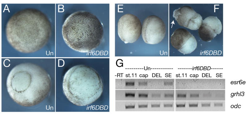
A) Animal and, C) vegetal views of stage 11 uninjected controls (Un). B) Animal and D) vegetal views of stage 11 embryos injected with 2 ng D. rerio irf6DBD mRNA. E) Uninjected (Un) and F) irf6DBD-injected stage 14 embryos. Arrow in F) indicates lesion in the ectoderm. G) RT-PCR analysis of SE markers, esr6e and grhl3 in control (Un) and irf6DBD-injected embryos and explants. St. 11, whole embryo stage 11; cap, stage 11 animal cap; DEL, deep layer explants; SE, superficial layer explants.