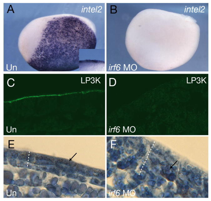
Figure 8. Maternal Irf6 is required for SE differentiation in X. laevis.

A,B) Whole mount in situ hybridization of stage 14, control A) uninjected (Un) and B) Irf6-depleted (irf6 MO) embryos for intelectin 2 (intel2). Lateral view is shown, anterior is to the left. Inset shows close up views of the epidermis in a bisected embryo. C,D) Sections of C) uninjected and D) Irf6-depleted stage 13 embryos stained with mAb LP3K. E,F) Sections of, E) control and, F) Irf6-depleted stage 22 embryos stained with Azan. Dotted lines indicate the thickness of the ectoderm; arrows indicate the location of embryonic pigment.
