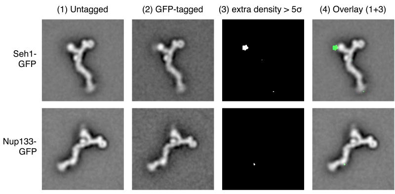
Figure 5.
Mapping of nup localization. Heptameric complexes were purified from yeast strains in which one protein of the subcomplex was genomically tagged with green fluorescent protein (GFP): the C terminus of Seh1 (first row), or the C terminus of Nup133 (second row). Aligned class averages of untagged and GFP-tagged particles are shown in columns (1) and (2). The significance map column (3) shows extra density for the GFP-tagged particles above the five-fold pixel-based standard deviation of the class averages. Column (4) shows an overlay of columns (1) and (3).

