
FIG 4.
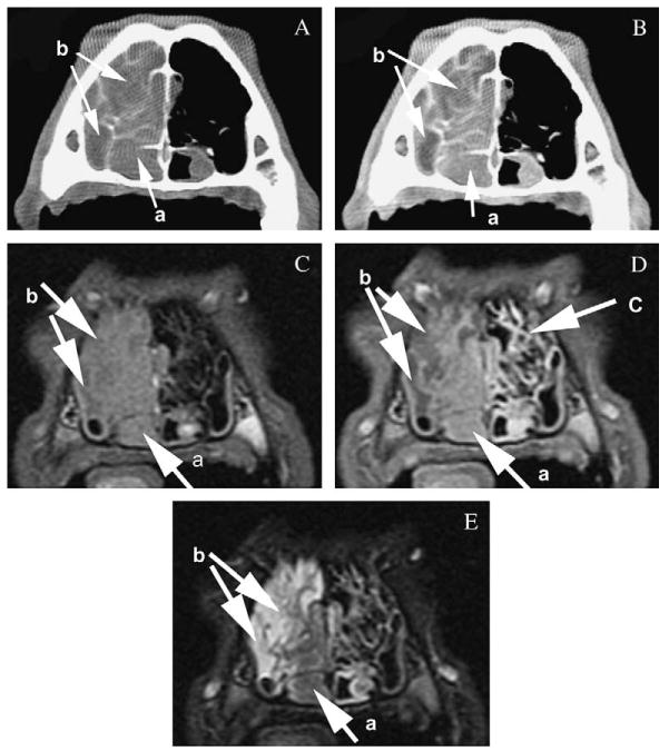
Comparison of the conspicuity of the features of canine nasal neoplasia on computed tomography precontrast (A) and postcontrast (B) images displayed in a soft tissue window and magnetic resonance imaging (MRI) T1-weighted precontrast (C), postcontrast (D) and T2-weighted (E) images at the level of the maxillary recess. Note the similar visibility of the neoplasia (a) based on enhancement on the postcontrast images and the depiction of fluid (b) based on non-enhancement or hyperintensity on T2-weighted images. The mucosal lining of the turbinates (c) is seen on T1-weighted postcontrast MRI images because of increased enhancement; however, the bony structure of the turbinates is not clearly identified