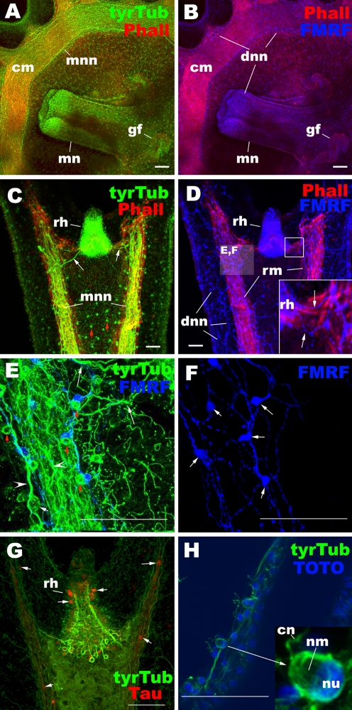
Fig. 2.
Ephyra neuromuscular system. Confocal sections of Aurelia sp. 1 ephyrae labeled with antibodies against tyrosinated tubulin (tyrTub), taurine (Tau), and/or FMRFamide (FMRF). In a–d, radial (rm) and circular (cm) muscle fibers are labeled with phalloidin (Pha). In h, nuclei (nu) are labeled with the fluorescent dye TOTO. In all subpanels, ephyrae are viewed from the oral side: a, b manubrium and surrounding subumbrellar epithelium of the bell, c–h rhopalar arm. The tyrTub antibody strongly labeled the motor nerve net (MNN) (a, c, e), which contains large bipolar neurons (white arrow in e) with longitudinally oriented thick neuronal processes (arrowheads in e). TyrTub-IR cnidocytes with apically located region devoid of staining, presumably occupied by nematocysts (nm), are also seen in the ectoderm (red arrow in c, e; inset in h). The taurine antibody labeled a subset of MNN neurons and sensory cells in the rhopalium (rh) (arrows in g). The FMRFamide antibody labeled the diffuse nerve net (DNN) (b, d–f), which contains multipolar neurons with thin neuronal processes (arrows in f). The tyrTub-IR cnidocytes lie alongside FMRFamide-IR DNN neuronal cell bodies and neurites (see red arrows in e), potentially indicating the presence of nervous communication. cm circular muscle, mn manubrium, gf gastric filaments, mnn motor nerve net, dnn diffused nerve net, rh rhopalium, cn cnidocil, nm nematocyst, nu nucleus

