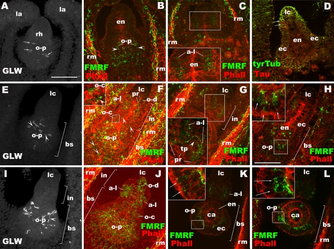
Fig. 6.
Development of rhopalial neurons outside of mechanoreceptors and ocelli in Aurelia sp. 1. a–d Prephyra I. e–h Prephyra II. i–l Prephyra III. Confocal sections of rhopalia labeled with antibodies against tyrosinated tubulin (tyrTub), taurine (Tau), GLWamide (GLW), and/or FMRFamide (FMRF). Phalloidin (Pha) is used to label filamentous actin. In all subpanels, specimens were imaged from the oral side. a Sections through the rhopalium, showing the oral-proximal group (o-p) of GLWamide-IR sensory cell bodies without basal processes (arrows). b Sections through the oral-medial portion of the rhopalium, showing FMRFamide-IR oral-proximal neurons (o-p) and their neurites (arrowheads). c A single section at the aboral-medial level, showing the presence of FMRFamide-IR aboral-lateral cells (a-l) without clear basal processes (inset). d A single section at the medial level, showing taurine-IR cells at the base of the ectoderm at the lateral side near the differentiating lithocyst (lc) (arrows). e Sections through the rhopalium, showing the formation of GLWamide-IR neurites (arrowheads). f Sections through the rhopalium, showing the oral-distal (o-d), aborallateral (a-l), oral-central (o-c; inset) and oral-proximal (o-p) groups of FMRFamide-IR cells and longitudinally oriented neuronal processes (arrowheads) connecting each of the clusters and the DNN outside of the rhopalium. g Sections at the level of FMRFamide-IR aboral-lateral sensory cells, showing the development of apical phalloidin-positive rings (pr) and their basal processes (arrowhead) forming commissure-like structures at the base of the touch plate (tp). h A single section at the medial level, showing FMRFamide-IR oral-proximal neurons (arrows). i Sections through the rhopalium, showing the oral-proximal group of GLWamide-IR sensory cells. Note the presence of a longitudinally oriented basal process (arrowhead). j Sections through the rhopalium, showing all major FMRFamide-IR neurons. k A single section at the aboral-medial level, showing FMRFamide-IR oral-proximal sensory cells (arrows). l A single section at the aboral-lateral level, showing FMRFamide-IR oral-proximal ganglion cells (arrows). Note their subepidermal position. rh rhopalium, la lappet, rm radial muscle, o-p oral-proximal neuron, a-l aborallateral neuron, en endoderm, ec ectoderm, in intermediate segment, bs basal segment, lc lithocyst, pr phalloidin-positive ring, o-d oral-distal neuron, o-c oral-central neuron, tp touch plate, ca rhopalar canal

