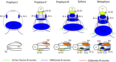
Fig. 8.
Schematic representation of rhopalial development from the prephyra I to the metephyra stages in Aurelia sp. 1. Upper row is oral view, and lower row is lateral view of the rhopalium. Prephyra I: statoliths, aboral-lateral and oral-proximal groups of neurons differentiate. Prephyra II: oral-distal, oral-central groups of neurons, as well as phalloidin-positive rings in areas of the future touch plate and the oral-distal group begin to develop. Prephyra III: tyrTub-/taurine-IR axons make connections with the MNN, and strong Taurine-immunoreactivity occurs in the touch plate cells, as well as in the oral-distal sensory cells. Ephyra: pigment-cup ocellus develops. Metephyra: pigment-spot ocellus develops. SL statolith, LC lithocyst, EN endoderm, O-D oral-distal group, A-L aboral-lateral group, O-C oral-central group, O-P oral-proximal group, SO spot-ocellus, TP touch plate, CO cup-ocellus

