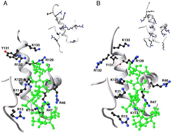
Fig. 5. Close-up of the pentasaccharide-binding site of native and latent antithrombin forms.
The image shows a close-up of the D- and P-helices and parts of the N-terminal region and the A-helix of the heparin-binding site of native antithrombin (A) and latent antithrombin (B) in complex with a high-affinity pentasaccharide (large pictures; PDB code 1e03) and in the unbound state (inset pictures; PDB code 1e05). A ribbon presentation of the selected parts of the protein backbone is shown in grey. The amino acid side chains of the residues of the N-terminus and the A- D- and P-helices that are known to participate in the interaction of pentasaccharide with native antithrombin are shown. Additionally, the amino acid side chains of Arg132 and Lys133, forming part of the extended heparin binding site of native antithrombin, and Tyr131, are shown. Carbon atoms of amino acid side-chains are drawn in black, nitrogen atoms in blue and oxygen atoms in red. The pentasaccharide is drawn in green. The images were produced in swiss PDB viewer.

