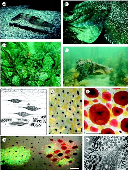
Figure 1.
(a) Iridescent spots in the squid Loligo pealeii. (b) Blue–green iridescence and white scattering leucophore stripes in cuttlefish (Sepia apama). (c) Camouflaged S. apama with pink iridescent arms and white markings caused by leucophores. (d) White leucophores in S. apama. (e) Skin in cross section showing the location of chromatophores (ch.) and structural reflectors (ir., iridophores; leuc., leucophores) in cephalopods. (f) Close-up of cuttlefish skin (Sepia officinalis) showing chromatophores (yellow, expanded; dark brown, partially retracted; orange, retracted) and white leucophores. Scale bar, 1 mm. (g) Brown, red and yellow chromatophores of squid (L. pealeii). Scale bar, 1 mm. (h) Combination of chromatophores and iridophores to illustrate the range of colours. Scale bar, 1 mm. (i) Electron micrograph showing iridophore plates (ir.) and spherical leucophores (leuc.) of cuttlefish (S. officinalis) skin. Scale bar, 1 μm (image courtesy of Alan Kuzirian).

