Figure 1.
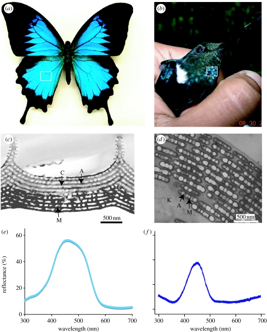
(a) The butterfly P. ulysses. (b) The hummingbird C. prunellei (photo by Juan Parra). In both cases, the box indicates the coloured region from which the transmission electron microscope (TEM) images and spectral data were taken. (c) TEM image of an iridescent blue scale from the butterfly P. ulysses, showing chitin (C) and air (A) arrays. The lower portion is much more darkly stained, suggesting the presence of diffuse melanin (M). (d) TEM image of an iridescent green barbule from the hummingbird Coeligena iris, showing highly ordered layers of air (A) filled melanin (M) platelets in a keratin (K) matrix. (e,f) Spectral data from P. ulysses and C. prunellei.

