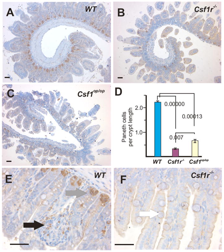
Figure 2. Lysozyme staining reveals a dramatic reduction of PCs in the SI crypts of two-week-old Csf1r−/− and Csf1op/op mice.
Low power images of lysozyme+ Paneth cells in the SIs of (A) WT, (B) Csf1r−/− and (C) Csf1op/op mice. (D) Quantitation of the number of lysozyme+ cells/crypt section (mean ± SEM, n = 3, ANOVA P-values shown), revealed significant differences the number of PCs at the crypt base (grey arrow in E) between both mutant and WT, as well as between mutants. (E) Higher power images of WT crypts showing the presence of lysozyme+ cells within the lamina propria indicative of macrophages (black arrow). (F) Only background lysozyme staining is evident in the Csf1r−/− SI. White arrow indicates goblet cell with enlarged mucin body. Bar = 50μm.

