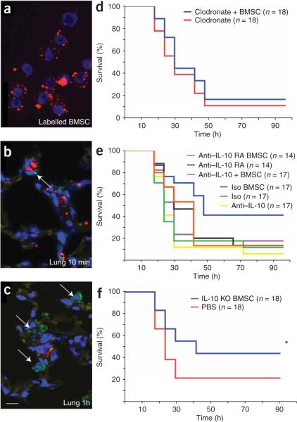
Figure 2.
Fate of injected BMSCs and effect of BMSC treatment on survival of normal and immune cell–depleted mice. (a–c) Immunohistochemical staining showing that BMSCs prelabeled with Q-dot (red punctate staining; a) travel to the lung (b) and take up residence in close proximity to macrophages (c). The latter cells were immunostained with an antibody to Iba1 (ionized calcium-binding adaptor molecule-1, a specific marker of the macrophage lineage47) and visualized with Alexa-Fluor-488 conjugated to a secondary antibody (green). Scale bar, 10 μm. (d–f) Summary of the effectiveness of BMSC treatment of mice genetically lacking or depleted of certain subsets of immune cells or soluble mediators. Survival curves show survival percentage of macrophage-depleted mice with or without BMSC treatment (d), survival percentage of BMSC-treated CLP mice and untreated mice after neutralizing IL-10 or blocking the IL-10 receptor (e) and survival percentage of after treatment with BMSCs derived from Il10−/− septic mice (f). *P < 0.05.

