Figure 5.
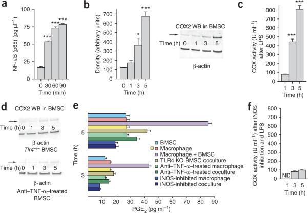
Studies of molecular alterations underlying the effect of BMSCs on macrophages. (a) NF-κB abundance in nuclei isolated from triplicate samples of BMSCs 30, 60 and 90 min after addition of LPS to the culture medium. (b) Western blot (WB) analysis of COX2 abundance in LPS-stimulated cocultures. Density measurements of three western blots are quantified in the bar graph. (c) The COX2 activity after LPS treatment of cocultures (triplicate samples). (d) Western blot analysis of COX2 abundance in Tlr4−/− (KO) BMSCs or in cultures treated with antibody to TNF-α (anti–TNFα). (e) Prostaglandin E2 (PGE2) abundance in macrophage cultures or coculture supernatants in a variety of conditions 3 and 5 h after LPS stimulation. Four samples were run for each condition. (f) COX2 enzyme activity after iNOS inhibition and 1, 3 and 5 h after LPS stimulation in triplicate samples. Error bars represent means ± s.e.m. *P < 0.05, ***P < .001.

