
Fig. 5.
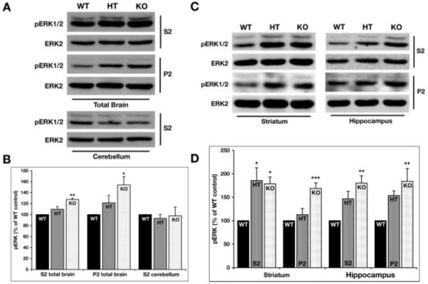
Increased baseline levels of phosphorylated ERK1/2 in STEP KO mice. (A) The levels of phosphorylated ERK1/2 were detected in the S2 and P2 fractions from total brain homogenates as well as the cerebellar S2 samples by probing with an antibody that recognizes ERK1/2 dually phosphorylated at the regulatory threonine and tyrosine residues (TPEYP-ERK1/2 or pERK1/2). The membranes were reprobed with anti-ERK2 antibody to ensure equal protein loading in each lane. STEP KO mice shows elevated levels of phosphorylated ERK1/2 when compared with WT controls in both the S2 and P2 fractions obtained from total brain homogenates. (B) Quantification of immunoblots for phosphorylated ERK2 expressed in the three genotypes normalized with total ERK2 protein loaded in the same blot (*P < 0.05 and **P < 0.01; n = 4). (C) pERK1/2 and ERK2 levels were detected in the S2 and P2 fractions from the striatum and hippocampus of WT, HT, and KO mice. The pERK1/2 levels were significantly elevated in both the fractions and regions tested. The HT showed significant increases only in the S2 fraction from the striatum. (D) The pERK1/2 levels from the immunoblots were quantified and normalized over the total ERK2 levels. The normalized pERK1/2 levels are plotted as a percentage of WT levels. The data were compared using one-way ANOVA, followed by posthoc Tukey HSD (*P < 0.05; **P < 0.01; ***P < 0.001; n = 4).