
Fig. 7.
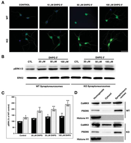
Enhanced phosphorylation of ERK1/2 in the KO hippocampal cultures and synaptoneurosomes. (A) The STEP WT and KO cultures were stimulated with increasing concentration of DHPG for 5 min and stained with pERK1/2 antibody. DHPG leads to activation of pERK1/2 in both the WT and KO cultures. However, the phosphorylation of ERK1/2 is more pronounced in the KO cells when compared with WT. This suggests that in the absence of STEP, there is a higher level of pERK1/2 activation [Scale bar-50 μm]. (B) Synaptoneurosomes prepared from WT and KO mice hippocampi were stimulated with increasing concentration of DHPG and processed for Western blotting. The membranes were probed with pERK1/2 and ERK2 antibodies sequentially. (C) The pERK1/2 levels from WT and KO synaptoneurosomes were measured using ImageJ and normalized to total ERK2 levels. The normalized pERK1/2 levels are plotted as a percentage of WT control levels. The data were compared using one-way ANOVA, followed by posthoc Tukey HSD (WT 20 μm: 126% ± 2%, P > 0.1; WT 50 μm: 138% ± 7%, P < 0.02; WT 100 μm: 143% ± 12%, P < 0.01; KO Ctl: 132% ± 5%, P < 0.02; KO 20 μm: 185% ± 19%, P < 0.01; KO 50 μm: 198% ± 13%, P < 0.005; and KO 100 μm: 237% ± 37%, P < 0.001; *P < 0.05; **P < 0.01; ***P < 0.001; n = 3). (D) The integrity of the synaptoneurosomes was determined by probing the input, cytosolic and synaptosomal fractions with histone H1 (present only in the input), CaMKII (detected in all fractions), and PSD95 (present mainly in the input and synaptosomal fraction).