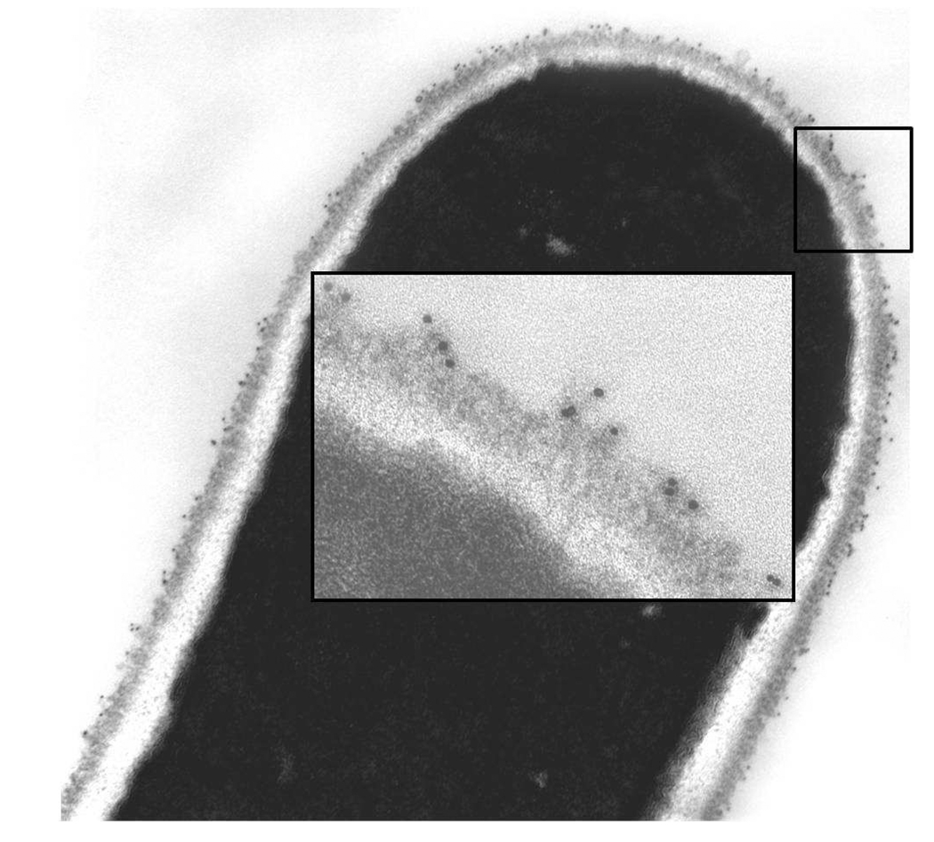
Fig. 6.
C. albicans CAI12 germ tubes grown in RPMI at 37°C for 45 min labeled with anti-Als3 113 MAb and a gold-conjugated secondary antibody. Gold particles were visible along the length of the germ tube. The inset shows a higher magnification image to visualize gold particles on the outermost layer of the C. albicans cell surface.

