Figure 6.

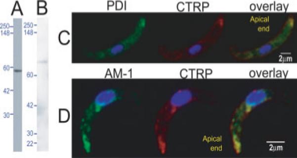
PDI and A-M1 locate at micronemes in ookinetes: ookinete lysate probed with (A) anti-PfPDI detects a protein of 51 kDa as expected for PDI and (B) anti-PfA-M1 (MAP1) detects two bands at 68 and 40 kDa, likely processed forms of PbA-M1, predicted to be 123 kDa. IFA of ookinetes probed with (C) PDI (green) co-stained with CTRP (red) and (D) A-M1 (green) co-labelled with CTRP (red). AM-1 and PDI clearly locate at the apical end of ookinetes and co-localise with CTRP. Additional perinuclear localisation of A-M1 and PDI is likely to be the ER and Golgi.
