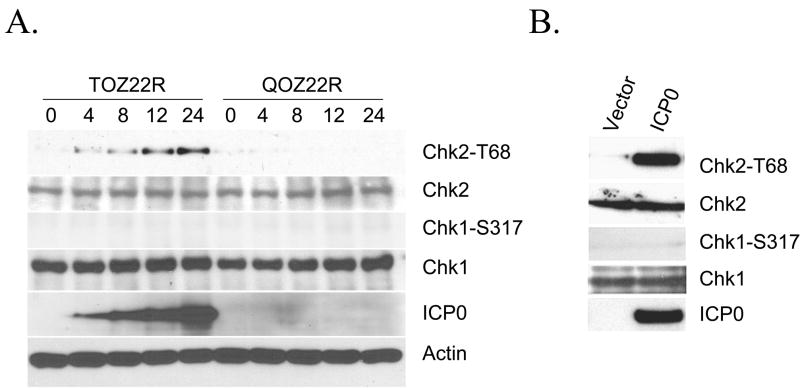
Fig. 2.
HSV-1 ICP0 activation of Chk2 kinase. (A) 293T cells were infected at an MOI of 2 with TOZ22R or QOZ22R and harvested at 0–24 hours post-infection (hpi). Cell lysates were electrophoresed and examined by immunoblotting with the indicated general or phospho-specific antibodies. The lanes are labeled by time point (hpi). (B) 293T cells were transfected with plasmid E110 expressing ICP0 or with empty plasmid. Cells were harvested 24 h later and processed for immunoblotting with the indicated antibodies.

