Figure 5.
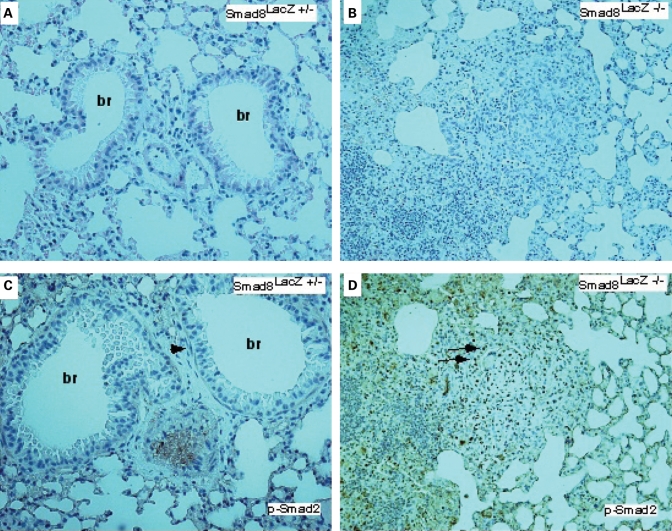
Expanded Activin/Tgfβ-signaling in Smad8 mutant pulmonary vessels. (A and B) H&E staining of control and Smad 8 mutant pulmonary vessels. (C and D) P-Smad2 immunostaining on control and Smad8 mutant. The arrow in (C) shows P-Smad2 immunostaining in a smooth muscle cell surrounding a bronchus. In (D), the Smad8 mutant has extensive P-Smad2 positive cells in an abnormal vascular lesion (all sections imaged at 200× magnification). br, bronchus.

