Figure 8.
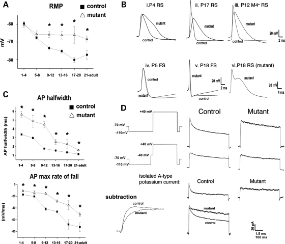
Hypomethylated neurons exhibit defects in resting membrane and action potentials. (A) Significant changes in resting membrane potential (RMP) were graphed (*P < 0.05, student t-test). (B) Action potential wave forms in control and mutant cells including M4-eGFP-positive neurons. (C) Action potential half-width and maximum rate of repolarization in P1–4, 5–8, 9–12, 13–16, 17–20 and P21–adult regular spiking cortical neurons in control and mutant slices. Note the increased half-width resulting from decrease in the maximal rate of repolarization at all ages. (D) Voltage schematic and leak subtracted currents elicited by stepping from −110 mV to +40 mV, with or without a 50 ms pre-step to −40 mV in outside out patches pulled from control and mutant RS cortical neurons. Figures are averages of currents obtained from 23 control patches and 8 mutant patches. Subtraction of pulses with the pre-step from pulses without the pre-step result in isolation of the rapidly inactivating potassium conductance (IA) in control neurons but not mutant neurons. Note that potassium currents in mutant somatic outside out patches inactivate much more slowly than those obtained from control neurons.

