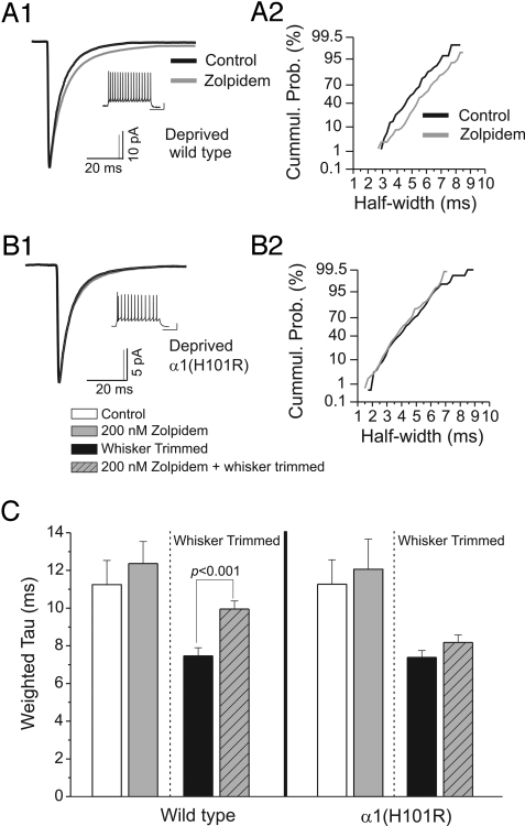
Fig. 4.
Inhibitory circuit remodeling depends upon increased α1 subunit function. (A1) Averaged inhibitory postsynaptic current (IPSC) traces from a whisker-deprived wild-type C57BL/6J mouse in artificial cerebral spinal fluid (ACSF; black, n = 81 events) and in zolpidem–ACSF (gray, n = 75 events). (A2) Cumulative distribution plot of IPSC half-width (using a nonlinear scale for the y axis) from the cell in A1 during ACSF (black) and zolpidem application (gray). (B1) Averaged IPSC traces from a deprived α1(H101R) knockin mouse in ACSF (black, n = 206 events) and during zolpidem (gray, n = 166 events). (B2) Cumulative distribution plot of IPSC half-width from cell in B1 during ACSF (black) and zolpidem application (gray). (C) Histogram of pooled values of τd,w derived from fitted IPSCs from low-threshold-spiking cells recorded from wild-type (Left) and α1(H101R) (Right) knockins. The hatched lines separate nondeprived from whisker-trimmed mice. Note that the deprivation-induced enhancement of IPSC decay in wild-type mice is eliminated in α1(H101R) mice.

