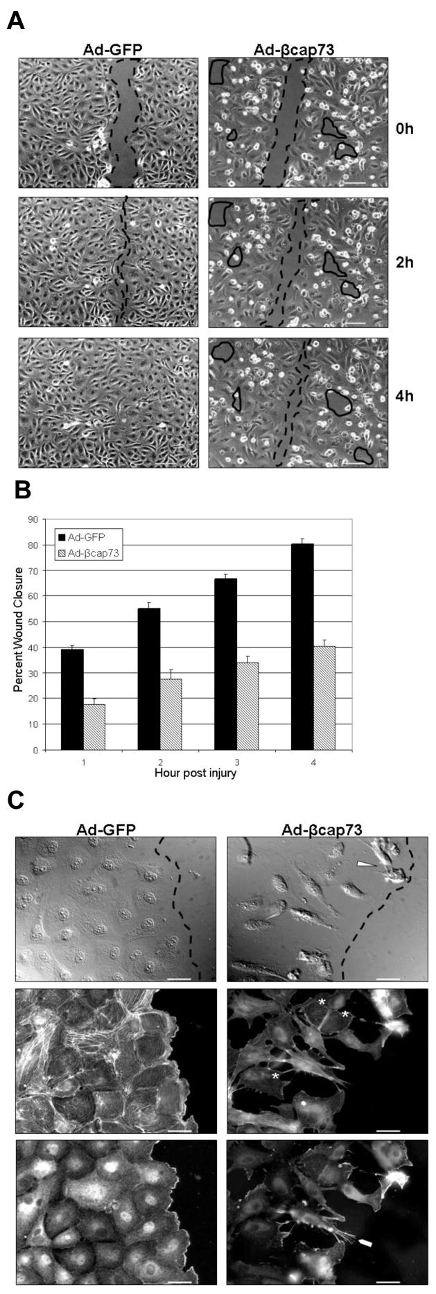
Figure 2. Ad-βcap73 inhibits the endothelial migratory response to injury.
(A) cEC monolayers infected with Ad-GFP or Ad-βcap73 were mechanically injured and migration was captured and quantified using digital imaging microscopy. In GFP-infected monolayers, post-injury migration persists unimpeded and wound closure is complete within four hours (injured space highlighted with dashed lines). However, in the βcap73 -overexpressing endothelial cultures, the wound remains unclosed due to defective cell shape and motility, which results in an imperfect monolayer where post-injury migration, cell-cell and cell-matrix contacts are perturbed (solid enclosed lines). Scale bar: 100μm (B) Quantitative analysis demonstrates that Ad-GFP wounds close steadily over the four hour experimental time period while the Ad-βcap73 wounds are impaired by approximately 50% in their healing rates. (C) cEC monolayers infected with either Ad-GFP or Ad-βcap73 were mechanically injured. Two hours post-injury, cells were processed for fluorescence imaging. In GFP-infected monolayers, cells polarize immediately post injury and establish a leading edge adjacent to the wound (dashed line). However, in βcap73 -infected cultures the contiguous monolayer is disrupted following perturbations in cell shape, motility, cell-cell and cell-matrix adhesions, which is apparent with differential interference contrast (DIC) imaging (gaps behind wound edge demarcated by dashed line), and following anti-β-actin IgG and fluorescent phalloidin localization. Note βcap73 -infected cells detaching from the matrix (arrowhead). Also, phalloidin-stress fiber staining is highly diminished in βcap73 -infected cells (asterisks) and β-actin rich filopodia (blunt arrowhead) are prevalent within the βcap73 -infected, but not control populations. Scale bar: 25μm

