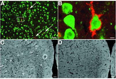
Figure 6.
AG-LI in rat cortex. Single, 14-μm coronal sections were single- or double-labeled with AG (red) and NeuN (green) antisera. (A) AG-LI associated with NeuN-positive cells (arrows). (Bar = 200 μm.) (B) The boxed portion of A at higher magnification projected from eight optical sections acquired at 1-μm intervals by using a ×60 objective. (Bar = 5 μm.) (C) AG-LI in cingulate cortex. (Bar = 50 μm.) (D) Absence of AG-LI in cingulate cortex after preabsorption with free AG (100 mM). (Bar = 50 μm.)

