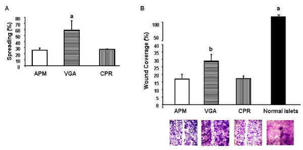
Figure 7.
Cell spreading and migratory ability. (A) Cells from the different human insulin-producing cell lines were seeded and allowed to adhere for 60 min. After removal of non adherent cells, the percentage of spread-out cells was determined at 45 min. Data were collected by random observation and at least in triplicate. a p < 0.001. ANOVA/Bonferroni's test. (B) Confluent monolayers of the human cell lines and of normal islets were "wounded". The cells were allowed to migrate into the cell-free area for 18 h and then photographed. Cell migration was evaluated by densitometry, calculating the area occupied by the migratory cells. Migration is expressed as the percentage (mean ± SD) of wound covered area. a p < 0.001 normal islets vs cell lines. b p < 0.001 VGA vs APM and CPR cells. ANOVA/Bonferroni's test.

