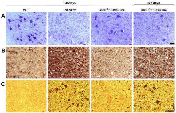
Figure 3.
Neuropathological and immunohistochemical studies of the anterior horn of the lumbar spinal cord. The first three columns show WTSOD1, G85Rflox, and G85Rflox/Lhx3:Cre mice at 345 days while the fourth column shows G85Rflox/Lhx3:Cre mice at 405 days with respect to Nissl staining (row A), GFAP immunoreactivity (row B), and SOD1 immunoreactivity (row C). The scale bar = 50μm

