
Figure 1.
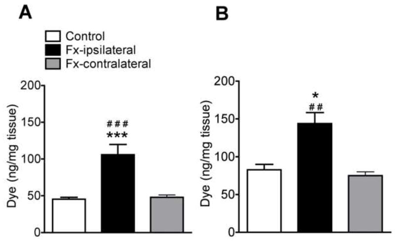
(A) Spontaneous dye extravasation 24 h after intravenous Evans blue dye injection. At 4 weeks after distal tibia fracture (Fx, n = 6) the dye content was increased by 133% in the ipsilateral, but not the contralateral hindpaw skin vs control rats (n = 7). (B) Intravenous substance P (SP) evoked dye extravasation in rats pretreated 5 min earlier with intravenous Evans blue dye. Five minutes after SP injection the hindpaw skin extravasation response was increased 73% in the ipsilateral, but not the contralateral hindpaw of fracture (n = 10) vs control rats (n = 5). All results are presented as the mean ± SEM in pg/mg wet tissue weight of the skin. *P < 0.05, and ***P < 0.001 for fracture vs control rat values, # P < 0.05, ## P < 0.01 for fracture vs contralateral hindpaw values.