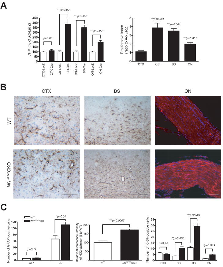
Figure 5. Neocortical astroglial cells exhibit no change in proliferation in response to Nf1 inactivation in vitro or in vivo.
(A) Whereas astroglial cells from CB, ON, and BS show increased proliferation in response to Nf1 inactivation, no change in CTX astroglial cell proliferation was observed following neurofibromin loss, as determined by [3H]-thymidine incorporation. (B) Increased numbers of GFAP-positive astrocytes are found in the CB and BS of Nf1GFAPCKO mice compared to controls. Similarly, increased NG2 fluorescence intensity in Nf1GFAPCKO mouse ON was observed relative to control mice. In contrast, no change in GFAP+ cell number was observed in the CTX of Nf1GFAPCKO mice compared to controls. Scale bars=100 μm. (C) Left, quantification of the number of GFAP-positive cells in the BS and CTX of Nf1GFAPCKO and control mice. Middle, quantitation of the relative NG2 fluorescence intensity in Nf1GFAPCKO and control mouse optic nerves. Right, Ki-67 (MIB-1) quantitation reveals significantly increased numbers of Ki-67+ cells in the CB, BS and ON of Nf1GFAPCKO mice compared to controls. No difference in the number of Ki67+ cells was observed in the CTX from Nf1GFAPCKO mice compared to controls. Error bars represent the mean±SEM.

