Fig. 5.
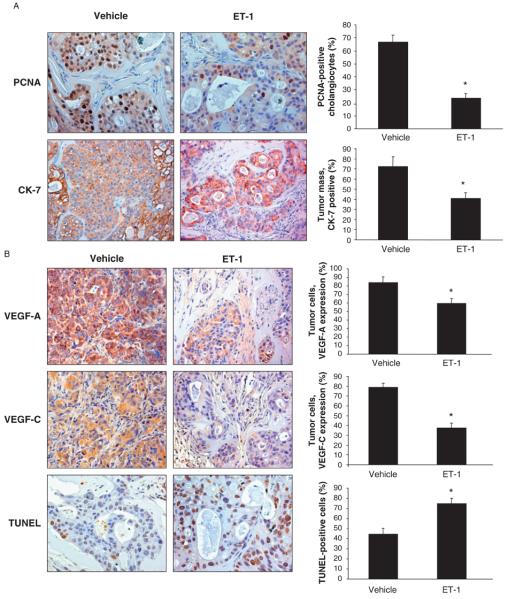
(A) Evaluation of proliferation [by staining for proliferating cellular nuclear antigen (PCNA)] in liver sections from tumour specimens from nude mice treated with endothelin-1 (ET-1) or vehicle. ET-1 decreased proliferation and increased apoptosis of cholangiocarcinoma cells in an in vivo mouse model. Data are mean ± SEM of three experiments. *P < 0.05 vs. PCNA-positive malignant cholangiocytes from vehicle-treated nude mice. Original magnification × 20. (B) Measurement of area occupied by cytokeratin-7 (CK-7)-positive malignant cholangiocytes from mice treated with ET-1 or vehicle. ET-1 administration significantly reduced the area occupied by CK-7-positive malignant cholangiocytes compared with mice treated with control solution. Data are mean ± SEM of three experiments. *P < 0.02 vs. CK-7-positive cells of tumour mass from vehicle-treated nude mice. Original magnification × 20. (B) Expression of vascular endothelial growth factor-A (VEGF-A) and VEGF-C by immunohistochemistry in tumours from ET-1- or vehicle-treated mice. Both VEGF-A and VEGF-C expression were reduced in malignant cells from ET-1- vs. vehicle-treated mice. Data are mean ± SEM of three experiments. *P < 0.05 vs. VEGF-A- and VEGF-C-positive tumour cells from vehicle-treated nude mice. Original magnification × 20. (B) Evaluation of apoptosis by terminal deoxynucleotide transferase end labelling (TUNEL) staining in tumours from ET-1- or vehicle-treated mice. An increase in the number of TUNEL-positive cholangiocytes demonstrated an increase in apoptosis in ET-1-treated tumours. Data are mean ± SEM of three experiments. *P < 0.02 vs. TUNEL-positive tumour cells from vehicle-treated nude mice. Negative controls (with pre-immune serum substituted for primary antibody) were also included. Original magnification × 20.

