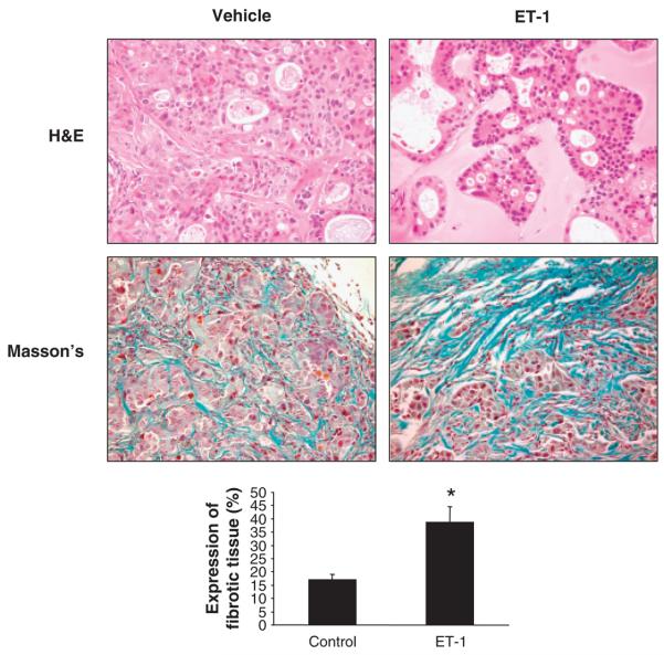
Fig. 6.
Photomicrographs of endothelin-1 (ET-1)- or vehicle-treated tumour sections stained with haematoxylin and eosin (H&E) and Masson's trichrome. Cholangiocarcinoma cells forming several acinar or tubular structures are shown in these images (H&E). An amorphous matrix localized between groups of cancer cells was observed in all the tumours. Masson's trichrome analysis showed fibrotic septa (blue) surrounding cholangiocarcinoma nodules. In ET-1-treated tumours, a wide area of fibrotic tissue is evident. Histological analysis demonstrated increased necrosis and damage and a more prominent fibrotic tissue in the ET-1-treated tumours with respect to controls. Original magnification × 80. Data are mean ± SEM of three experiments. *P < 0.05 vs. vehicle-treated tumour.

