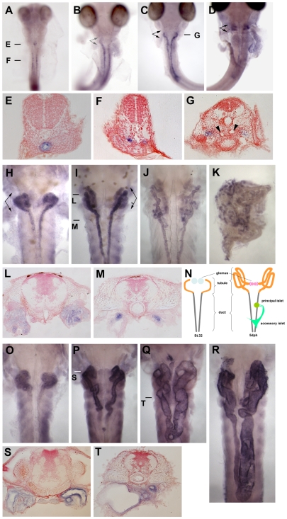
Figure 2. pc/glis3 expression in medaka kidney.
The expression pattern of pc/glis3 mRNA in embryonic kidney of wild-type orange red medaka is shown for larvae of (A) stage 27, (B) stage 30, (C) stage 32, and (D) stage 35. In sections (E) and (F) (stage 27) and (G) (stage 32), pc/glis3-positive cells can be observed in the epithelium of the gut, the renal duct, and the renal tubule, respectively. Arrowheads in (G) indicate the pronephric glomus. pc/glis3 expression in larval kidney of wild-type medaka is shown for (H) the hatching stage, (I) 5 days posthatching (dph), (J) 10 dph, (K) 20 dph, and (L, M) sections of the 5 dph fry in (I). In (N), segments of the kidney and the pancreas at stage 32 (left) and at 5 dph (right), are shown [33]. At stage 32, developing pronephric glomera (blue) are located most anteriorly, followed by the pronephric tubules (orange) and ducts (gray). The pronephric tubules and glomera develop after the pronephric ducts have formed [33]. By 5 dph, the pronephric glomera (pink) and tubules (orange) have become mature. The pancreatic principal (green) and accessory (dark green) islets can be visualized by in situ hybridization with insulin. Expression of mutant pc/glis3 mRNA in pc mutant kidney is shown for (O) the hatching stage, (P, Q) 5 dph, (R) 10 dph, and (S, T) 5 dph. The pc mutant individual shown in (Q) and (T) has more severe dilation of the renal tubules and ducts than that in (P) and (S). Arrows in (B), (C), (D), (H), and (I) indicate the positions of tubular segments. Bars in (A), (C), (I), (P) and (Q) indicate the positions of the sections shown in (E), (F), (G), (L), (M), (S) and (T). The sections were stained with neutral red to visualize the cell nuclei.

