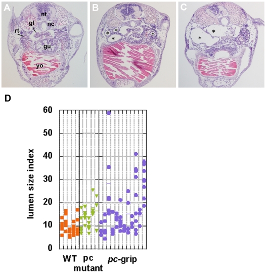
Figure 3. Cyst formation in pc/glis3 knockdown individuals.
Gene-specific knockdown with antisense oligonucleotides resulted in dilation of the renal tubules (B), ducts (not seen in this figure), and glomera (C). In (A), a cross section of a hatching fry with no notable dilation is shown. gl, glomus; gu, gut; nc, notochord; nt, neural tube; rt, renal tubule; yo, yolk. Typical dilation of the tubule is indicated by a star (B, C). The size of the lumen of the renal tubule was measured and is presented as a lumen size index (D). The lumen size index was defined as being a perpendicular bisector of the greatest tubule diameter of the lumen in a section. This was done in order not to overestimate the lumen size in the event that the lumen was sectioned at a right angle or the lumen shape was oval. Mean lumen size index values are indicated by a horizontal bar and measurements were taken for several sections of the tubule (arrows in Fig. 2H) at locations where the glomus was observed in the hatchlings. The lumen size indices for each individual are shown in a single lane, with individual dots in each lane representing the lumen size index value of a single tubular section. Results from knockdown with pc-grip (pc-SPD) in wild-type medaka as well as from pc mutants are shown. Compared to untreated wild-type medaka, the lumen size index in pc/glis3-knockdown fry is significantly higher (p<0.005).

