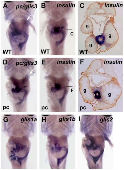
Figure 4. pc/glis3 expression in the medaka pancreas.
In situ hybridization was performed using 5-day posthatching fry with intact internal organs. Compared with Fig. 2N, the principal and accessory islets of the pancreas can be easily seen to be positive for the expression of all genes shown. The pancreas often varies in shape. White arrows indicate staining in the renal tubules. Arrowheads indicate the principal islets. (A, B, C) wild-type. (D, E, F) pc mutant. (A, D) pc/glis3, (B, C, E, F) insulin, (G) glis1a, (H) glis1b, and (I) glis2 expression was detected. (A, B, D, E, G, H, I) ventral view. The principal islet sections show that there is no apparent difference in the number of insulin-positive cells (C, F).

